Killing germs with electron beams
Medical devices, packaging and food can be safely and efficiently sterilized using electron beams. In the future, Fraunhofer researchers also want to use accelerated electrons to remove germs from tissue transplants and also change the properties of the biological material.

Medical needles, disposable syringes and dialysis tubes, but also pharmaceutical packaging must be germ-free. Electron beam sterilization is a proven method for killing bacteria, viruses, fungi, spores and the like. This is done by accelerated, high-energy electrons that penetrate the material. Researchers at the Fraunhofer Institute for Organic Electronics, Electron Beam and Plasma Technology FEP in Dresden successfully use this method, for example, to rid seeds of pests. Within milliseconds, the DNA of the pests is destroyed, thus disabling their ability to reproduce (see link below). Now the scientists are transferring their expertise to new areas of application: they are investigating the possibilities of non-thermal electron beam technology for sterilizing and modifying biological tissue. This opens up new treatment options in the medical field.
Advantage: fabric does not heat up
"We treat tissue samples under atmospheric pressure and at temperatures below 40 degrees Celsius with electrons that are only accelerated just enough to penetrate the material. This is called low-energy electron beam treatment," explains Dr. Jessy Schönfelder, head of the Medical Applications group at FEP. The advantages of this method, which is gentle on the material, are that the tissue does not heat up, cells remain intact - the depth of action can be adjusted and thus only the outer surface layer can be sterilized. With REAMODE (Reactive Modification with Electrons), the scientists at FEP are using a research facility that can be adapted to a wide variety of applications of low-energy electron beam technology. Parameters such as penetration depth and intensity can be specifically determined.
Crosslinking tissue with accelerated electrons
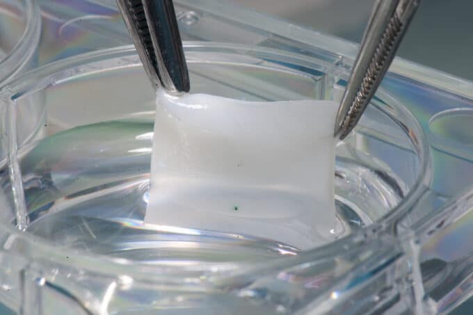
Using accelerated electrons, the researchers can also specifically influence material properties and bring about changes in tissue. For their experiments, they chose the pig pericardium as a biological model. Physicians use this tissue as a biological heart valve replacement. However, the implants last a maximum of 15 years. The reason: the chemical glutaraldehyde used for the necessary cross-linking of the tissue also causes calcification of the biological implants in the medium term. "Accelerated electrons are an interesting alternative here due to their property of splitting chemical bonds and enabling cross-linking," Schönfelder explains. In their experiments, the researcher and her team were able to demonstrate cross-linking of collagen molecules, i.e. protein chains.
To prove the cell compatibility of electron beam sterilization, the researchers seeded cell cultures on different samples. Only a small proportion of cells grew on the tissue treated with the toxic glutaraldehyde. On the electron-beam-modified counterpart, on the other hand, significantly more cells grew and were able to proliferate. In comparison, even fewer cells settled on untreated samples than on the electron-treated samples.
Cell functions are preserved
Tests with biological vascular prostheses - for which the experts used aortas from animal models - were also successful. "In patients with blood vessel diseases, the replacement of vessels with synthetic prostheses made of polymers is unavoidable. These are limited to diameters of six millimetres or more due to the risk of thrombosis. Biological implants have to be used for vessel diameters smaller than six millimetres," said Schönfelder. One problem is their sterilisation. The muscle and endothelial cells in the inner layers of the blood vessels must not be damaged. Since the researchers can use their equipment to determine the optimal penetration depth of the electrons into the vessel wall and only sterilize the outer layer of the aorta, the cells retain their functionality. The penetration depth in the tests was 23 micrometers, which is within the outermost connective tissue layer. "At a radiation dose of 22 kilogray (kGy), vascular functions were not affected. At the same time, the bacteria applied to the sample were killed safely and within a few seconds," confirms the chemist. The low acceleration energy enables compact designs of the devices for electron treatment. Due to the simultaneously high treatment speed, the method is predestined for use in a tissue bank or in the operating theatre. Now the experts want to optimize the process and, in the next step, build a sterilization device adapted to the method.
Source: Fraunhofer FEP


